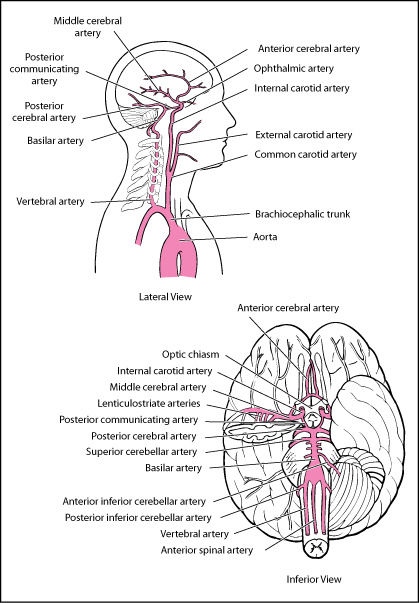
Nutrients and oxygen are transported to the highly metabolic brain by means of an efficient circulatory system. The cerebral blood supply system is mainly dependent upon the two anterior cerebral arteries that supply oxygen to medial portions of the frontal lobes and the superior medial parietal lobes. An anterior communicating artery connects one anterior cerebral artery to another. These arteries contribute to the healthy and normal functioning of the brain.
Important Cerebral Arteries
Right and left vertebral arteries and common carotid arteries supply blood to the brain, scalp and face. Vertebrobasilar arteries supply brain stem and two fifths of cerebrum with blood. There are two types of common carotid arteries:
- External carotid arteries- the set of blood vessels that supply blood to scalp and the face.
- Internal carotid arteries- the set of blood vessels that supply blood to anterior three fifths of cerebrum excluding occipital and temporal lobes.
Inadequate blood flow through internal carotid arteries leads to impaired frontal lobes functioning. This may be due to weakness, numbness or paralysis of the side opposite the occluded artery. Occluded artery may lead to paralysis or blindness.
Vertebrobasilar and carotid arteries communicate in a circle at the base of the brain; this is called the Circle of Willis. Different arteries arise from this circle and travel to the various parts of the brain, these arteries may include:
- Middle cerebral artery (MCA)
- Anterior cerebral artery (ACA)
- Posterior cerebral artery (PCA)
As already established, vertebrobasilar and carotid arteries form a circle so even if any main artery gets obstructed, the distal smaller arteries that were previously getting blood from it can now receive from other arteries. This phenomenon is called collateral circulation.
Anterior Cerebral Artery
Location of ACA
Located on top of the brain, ACA serves the outermost surface of the cerebral hemisphere. This portion comprises of a long strip extending from frontal lobe to the brain’s back. This region also comprises of olfactory tract and bulb, anterior portions of basal ganglia and internal capsule, and a part of lateral surface of frontal and parietal lobe that is located next to the medial longitudinal fissure.
Branches of ACA
Anterior cerebral arteries branch into smaller arteries that are identified from A1-A5. A1 is connected to internal carotid artery from the base of its segment and stretches all the way to the anterior communicating artery. A2 stretches from anterior communicating artery to the callosomarginal and pericallosal arteries (A3). From segment A2, the frontopolar and orbitofrontal arteries extend as well. A3 comprises of internal parietal and precuneal arteries. Segment A4 and A5 are the small branches extending from the anterior cerebral arteries. These are often termed as callosal arteries.
Functional areas of brain that ACA supports
Anterior cerebral artery is responsible for supplying blood to the regions of frontal lobes and superior medial parietal lobes. Inadequate blood flow to the brain leads to sensory deficit, paralysis or stroke. ACA supply blood to the anterior part of the frontal lobe, which deals with the cognition of higher level including reasoning and judgment. Occluded arteries result in speech difficulties, apraxia of gait that may affect the arms movement and dementia. Apraxia is characterized as the loss of ability to carry out different functions that a person is physically able to perform otherwise. Apraxia of gait is associated with walking and the affected person walks with flat, short steps. Anterior cerebral artery rises from the internal carotid and stretches at a right angle, branching and supplying blood to different parts of the brain. It supplies blood to the following parts of the brain:
- The septal area: It deals with regulating pleasure response and fear.
- The corpus callosum: It’s a thickened fibrous band that is responsible for dividing the brain into two halves.
- The primary somatosensory cortexes for the leg and foot: These are the areas responsible for interpreting the sense of touch for leg and foot.
- Motor planning areas of the frontal lobes: The area of the brain that influences judgment and planning.
Anterior Cerebral Artery Occlusion
The Occlusion of ACA will lead to the compromised functioning of the corresponding areas of brain: basal ganglia,anterior corpus callosum ,anterior fornix and the medial aspects of the frontal as well as parietal lobes.
The occlusion may result from:
- Emboli, usually resulting from narrowing of artery due to atherosclerosis at the bifurcation of the carotid artery either from atheroma arising in the aortic arch or from cardiac source.
- Superimposed thrombosis in conjunction with atherosclerotic stenosis.
Lacunar strokes may result from lipohyalinotic intrinsic disease that that targets small penetrating vessels.
The internal carotid artery is most commonly occluded at the site of proximal 2 cm to the artery’s origin and intracranially occluded at the carotid siphon. The factors that may influence the degree of infarction may be the systemic blood pressure and the speed of occlusion.
Occlusion in the proximal segment of ACA will produce minor deficits thanks to the anterior communicating artery. But occlusions further away from this area will, on the other hand, result in severe ACA syndrome, the major symptoms of which include hemisensory loss of lower extremity and contralateral hemiparesis.
