
Ascites is a condition that is characterized by the accumulation of fluid in the abdominal region. Often, it occurs due to improper functioning of liver that leads to abnormal accumulation of fluid in the space present between the lining of the organs and the abdomen. However, people who are diagnosed with ascites are reported to only have a ranging rate of 30-40%survival after 5 years of initial diagnosis. If you think you are experiencing the symptoms associated with ascites then seek medical help right away. With an ascetic fluid analysis—as the key diagnostic for ascites, your doctor can better monitor your condition and offer the most effective treatment.
What Can Ascitic Fluid Analysis Include?
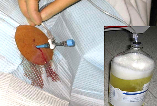
Ascitic fluid analysis or peritoneal fluid analysis is the major diagnostic test to study the pathophysiology of accumulation of fluid in the peritoneum, including diagnosing the causes and inflammation of the fluids. As for the fluid, the inflammatory collection is exudate, and the non-inflammatory collection is transudate. Some patients may wonder what tests are included. Read on to find out what to expect in the analysis.
1. Gross Appearance
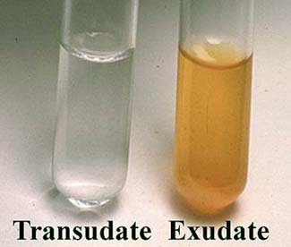 The transudate fluid can be straw colored and clear, yet the color may appear milky in blocked lymphatic vessels. This may particularly be seen in carcinoma, tuberculosis and lymphoma, which contains more than 100mg/dl triglycerides.
- The color of the fluid becomes opaque or turbid due to the presence of any inflammatory conditions such as appendicitis, pancreatitis and peritonitis.
- Abdominal bleeding or trauma, hemorrhagic pancreatitis or tumor infiltrate may result in hemorrhagic color.
- In case of intestinal perforation, pancreatitis or ruptured gall bladder may cause bile stained or green colored fluid.
2. Specific Gravity
The specific gravity is essential in the ascitic fluid analysis. The specific gravity of the exudate is greater than 1.015, yet the figure is less than 1.015 for transudate.
3. Proteins
The proteins present in exudate are more than 3g/dl but less than 3g/dl in transudate. In order to differentiate transudate from exudate, serum protein and ascitic fluid protein ratio is of great importance. The ratio's value must be greater than 0.5 for the diagnosis of exudate.
4. Cell Count
There are fewer than 250/µL polymorphonuclear leukocytes (PMNs) and fewer than 500/µL leukocytes in normal ascitic fluid. But, in an inflammatory condition, the white blood cell count will arise. If the PMN count increases to 250/µL or more, there are high chances of the presence peritonitis. For the tuberculosis and peritoneal carcinomatosis cases, lymphocytes are predominant.
In inflammatory conditions there is a greater number of reactive mesothelial and polys, whereas in case of transudate, there may be a greater number of lymphocytes. This may also be seen in malignant ascites.
5. Cytology
This test is better done on cytospin. It helps in differentiating as well as finding the malignant cells. At times it's difficult to differentiate between the malignant and mesothelial cells.The morphology of mesothelial cells differs with respect to cellular content and nuclei. There may be clumping of chromatin and altered ratio of nucleus and cytoplasm. Prominent nucleoli may be present.
6. Glucose
Glucose level is mostly equal to that of blood glucose level; however, it's low in case of bacterial ascites and tuberculosis.
7. Amylase
Amylase is elevated if conditions like pancreatic pseudocyst, intestinal perforation, intestinal necrosis, acute pancreatitis and pancreatic trauma are present.
8. Lactate Dehydrogenase
Also referred as LDH, it is a diagnostic test to calculate the exudate ratio between ascites LDH and serum LDH. The figure can be greater than 0.6 in exudate.
9. Gram Stain or Culture Test
Gram staining and culture test both can determine what type of bacteria is present in an infection. However, in ascitic analysis, the gram test isn't usually performed because mostly culture and sensitivity tests are preferred.
As for detecting the bacteria of the ascitic fluid, culture test has a ratio of sensitivity at 92% with samples being inoculated into blood culture bottles timely. While, gram staining procedure just has a ratio at 10% in detecting spontaneous bacterial peritonitis.
When Should You Take an Ascitic Fluid Analysis?
 Aside from being suspected to have ascites or peritonitis, the doctor will ask you to get ascitic fluid analysis done for the following conditions:
- Ascites having unknown origin
- Tenderness and abdominal pain
- Intestinal perforation
- Any suspected intra-abdominal malignancies
Possible Ascitic Fluid Analysis Results
1. Ascites with Transudate
When serum albumin/ ascites albumin ratio is greater than 1.1gm/dl, it may present with following clinical manifestations:
- Viral hepatitis in conjunction with massive or sub-massive liver necrosis
- Obstruction of inferior vena cava
- Constrictive pericarditis
- Meig's ovarian tumor syndrome
- Congestive heart failure
- Liver cirrhosis
- Budd Chiari syndrome (obstruction of hepatic vein) in conjunction with tumors like pancreatic cancer, hypernephroma and hepatoma, or in conjunction with hematological disorders like myeloid metaplasia, polycythemia vera and myeloproliferative disease or as a result of infections.
90% ascitic fluids are transudates that may result from cirrhosis or congestive heart failure. The ascitic fluid analysis may yield:
- Clear appearance of fluid
- Typically low serum-albumin ascites level; if the ratio is greater than 1.1g/dl then it confirms the presence of transudates.
- Presence of few cells
2. Ascites with Exudate
When serum albumin/ ascites albumin ratio is less than 1.1gm/dl, it may present with following clinical manifestations:
- Hemodialysis CRF associated ascites
- Previous abdominal trauma with ruptured lymphatics
- Amyloidosis
- Struma oovarii
- Pseudomyxoma peritonei
- Lymphatic obstruction which may be a lymphoma or intestinal lymphangiectasia
- Sarcoidosis
- Myxedema
- Transected lymphatics next to portal caval shunt surgery
- Post-surgery starch or talc peritonitis
- Pancreatitis
- Tuberculous peritonitis
- Peritoneum related neoplastic diseases like peritoneal carcinomatosis and lymphomatous disorders.
- The ascitic fluid analysis yields:
- Cloudy appearance of fluid
- High serum-ascites albumin level as compared to transudates i.e. <1.1 g/dl
- Increased cellular count
Based on the suspected cause of exudates, the following diagnostic tests may be recommended by your doctor:
- Test to assess the physical appearance: Normal color of peritoneal fluid is straw colored.If cloudy colored—presence of white blood cells or microorganisms; if reddish colored—presence of blood; if yellow colored—liver disease and may become milky due to lymphatic system obstruction; if greenish colored—presence of bile.
- Chemical tests: glucose, tumor markers and amylase is checked.
- Microscopic examination:elevated WBCs in malignant condition; elevated number of neutrophils in bacterial infections.
3. Disorders That Cause Ascites
- Obesity
- Mesenteric cyst
- Ovarian cyst
- Pancreatic pseudocyst
- Hydronephrosis