
Posterior horn of medial meniscus plays a vital role in the maintenance of normal knee functions. Besides performing highly specialized functions such as maintenance and distribution of weight among knee muscles, posterior horn of medial meniscus also works as the shock absorber in order to prevent serious injuries in situations of sudden trauma. However, it is imperative to keep in mind that the central position and key roles in the knee function support also increase the risk of injury to posterior medial horn of meniscus. Most common forms of injury include ruptured or a torn meniscus, which if left poorly managed, can aggravate the risk of musculoskeletal conditions like osteoarthritis.
What Is Posterior Horn Medial Meniscus?
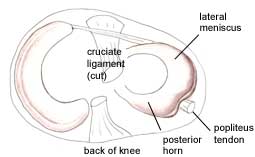 The posterior horn of medial meniscus is a part of medial meniscus that is situated along the posterior aspect of the knee and mainly serves as the primary weight bearing component of your medial meniscus. Medial meniscus is responsible for transmitting approximately 50% of your weight directed at the medial compartment of the lower limb. Posterior horn of medial meniscus provides the much needed shock absorbing capability to the meniscus in order to function appropriately.
What Is the Common Problem with Posterior Horn Medial Meniscus?
Posterior horn of medial meniscus is the primary weight-bearing component of the medial meniscus, so it is also a common site of tearing or injuries. The most susceptible population is individuals who are already suffering from ACL (or anterior cruciate ligament) tear, because in all such instances, posterior horn of medial meniscus becomes the key role in preventing the knee slippage in the anterior (forward) direction.
Following are some symptoms which are most commonly observed in patients with posterior horn medial meniscus tear:
- Knee stiffness
- Pain in the affected area is the most frequently reported complaint, which is often associated with swelling of knee and will become severe when deep squatting
- Foot muscle instability
- Locking of knee
How to Diagnose Posterior Horn Medial Meniscus Tear
There are multiple ways to diagnose this kind of tearing. Some of the important diagnostic methods are discussed below.
1. Patient History
The past medical history and family history of the patient provide significant information in diagnosing the disease. If any person in the patient’s family is suffering from the same problem, then a provisional diagnosis can be made easily.
2. Physical Examination
Healthcare providers perform physical examination after conducting a thorough history of the patient to assess swelling and tenderness over the affected knee. One of the diagnostic procedures conducted during physical examination is McMurray test, in which the physician bends the patient's knee and straightens it in the forward and backward direction. A tension over medial meniscus region can result in clear clicking sound and characteristic pain.
3. Imaging Tests
Two common imaging tests are performed to confirm the diagnosis:
- X rays: It is not categorized as one of the best tool for diagnosing the posterior horn meniscus tear, but it can rule out other causes associated with the pain, such as osteoarthritis.
- MRI (magnetic resonance imaging): MRI is a sensitive imaging test for diagnosis of posterior horn medial meniscus tear.
How to Treat Posterior Horn Medial Meniscus Tear
Treatment or management protocols for posterior horn menial meniscus tears are quite challenging. It is estimated that only 10% of the injuries involving the tear of posterior horn medial meniscus are repairable. The primary objective is to control the disease process to avoid the complications like osteoarthritis.
The patients are recommended to visit the clinic after regular interval of time, so small changes in symptoms can be noted down and any progression of disease can be identified well. Some of the treatment protocols can be planned according to the patient's condition which includes:
- Partial meniscectomy
- Physical therapy
- Meniscal transplantation
- Activity modification
Partial medial meniscectomy is a surgical procedure in which partial meniscus (including the torn portion) is removed via excision. Soon after the surgery, physical therapy is advised to be started to reactivate the surrounding muscles including quadriceps muscles. This will help in regaining the muscle mass and knee mobility in a short span of time.
Generally, patients, in whom small portions of meniscus are trimmed, can perform light activities until at least 6 weeks after the surgery. In addition, physical therapies should be continued during this interval.
In patients whose significant portion of posterior horn of medial meniscus is removed, stressful activities should be completely prohibited as there is a greater risk of developing osteoarthritis. Individuals are advised not to move their knee more than 90 degree during knee flexion. High impact activities should be avoided for at least 6 weeks after surgery.