
Next to heart disease, breast cancer is the leading cause of death in women all over the world. Although the exact cause of breast cancer is unclear, early detection and treatment saves lives and reduces cost. Fortunately, it is becoming easier to diagnose the disease through different technologies, with ultrasound imaging as one of the frequently used diagnostic tools.Here we show somebreast cancer ultrasound images and important facts for your information.
Breast Ultrasound
Ultrasound technology is an important way to diagnose various conditions. Sound waves from a device bounce off tissues, creating images that can help doctors visualize normal structures and abnormal growths. Ultrasound imaging of the breast helps detect different types of conditions, including non-cancerous (benign) and cancerous (malignant) lesions.
If a doctor suspects an abnormal growth in the breast after a clinical breast examination or screening mammography, evaluation of breast cancer ultrasound images will help confirm the diagnosis. One advantage of ultrasound technology is that it allows substantial freedom in obtaining breast images from any orientation.
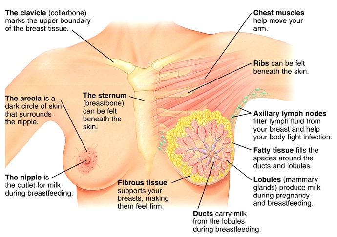
(Picture: Structure of the breast)
Breast Density
During a mammography, the density of breast tissues is determined. The images produced show white and gray areas, which distinguish the breast tissues and fat tissues, respectively. The amount of dense breast tissues differs in every woman, and experts usually describe the density levels in grades (1 to 4).
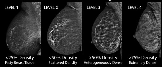
Generally, whiter mammogram images indicate denser breasts. Most women normally have grade 2-3 breast density. However, cancer can also be seen as white, making it a bit obscure in women with extremely dense breasts. This makes diagnosis a bit tricky using mammography, since up to half of all cancers may be missed in women who have very dense breasts. Studies also show that women with extremely dense breasts are about five times more likely to develop breast cancer than women who have level 1 breast density.
Common Types of Breast Cancer and Ultrasound Images
Some of the different types of breast cancer are named according to their origin.
Cancer Starting from Milk Ducts
The most common type of breast cancer is ductal carcinoma, which forms in the linings of the milk ducts within the breast. The milk ducts carry your breast milk from lobules, where milk is produced, to your nipple.
Cancer cells can remain within the milk ducts and this is considered as noninvasive cancer or ductal carcinoma in situ. However, cancer cells can also break out of the ducts and invade surrounding tissues (invasive ductal carcinoma). Here are some breast cancer ultrasound images:
Ductal Carcinoma in Situ (DCIS)
.jpg) DCIS is breast cancer that is non-invasive, where cancer cells are contained in the linings of the milk ducts.
DCIS is breast cancer that is non-invasive, where cancer cells are contained in the linings of the milk ducts.
Ductal carcinoma in situ is usually seen as linear microcalcifications (see arrows), which demonstrate linear orientation, as seen in this patient. This type of cancer precedes the development of a distinct mass, lump or invasive cancer. Mammography helps identify breast cancer in this early stage, which is one of its main benefits.
Invasive Ductal Carcinoma (IDC)
.jpg) Invasive breast cancer develops when abnormal cells originally found in the linings of the breast milk ducts invade surrounding tissue.
Invasive breast cancer develops when abnormal cells originally found in the linings of the breast milk ducts invade surrounding tissue.
A breast mass (arrows) is shown here with indistinct, spiculated borders, which suggest the presence of tissue invasion in invasive ductal carcinoma. This is the most common type of breast cancer.
Cancer Starts from Milk-producing Lobules
Cancer can also start from the lobules of the breast, where milk is formed. Lobular carcinoma can also break out and spread out, and this is known as invasive lobular carcinoma. Lobules connect to the ducts, which carry the milk to the nipples.
Invasive/Infiltrating Lobular Carcinoma (ILC)
.jpg) This breast cancer ultrasound image shows changes related to breast cancer that are not seen as microcalcifications or a mass or lump. Rather, the right breast is seen as smaller than the left breast. There is a slight increase in the density in the right breast compared with the left. You can also see some benign calcifications scattered in the breast tissues. Compared with invasive ductal carcinoma, invasive lobular carcinoma usually has a more subtle appearance, as shown in these images.
This breast cancer ultrasound image shows changes related to breast cancer that are not seen as microcalcifications or a mass or lump. Rather, the right breast is seen as smaller than the left breast. There is a slight increase in the density in the right breast compared with the left. You can also see some benign calcifications scattered in the breast tissues. Compared with invasive ductal carcinoma, invasive lobular carcinoma usually has a more subtle appearance, as shown in these images.
Inflammatory Breast Cancer (IBC)
 This is a less common type of breast cancer. This condition usually does not develop a lump, but commonly affects the breast skin.
Inflammatory breast carcinoma is a rare type of invasive ductal carcinoma, which mimics an infection or mastitis at presentation. Although the affected breast is not usually very tender, it is red, swollen, and warmer than the other breast. The skin over it is stretched and thick, looking like an orange peel. Enlargement of axillary lymph nodes is also common. On imaging, it is sometimes difficult to distinguish this condition from mastitis, although clinically, patients with mastitis are often in so much pain.
Watch this video to learn more about breast cancer ultrasound images: