
The wall that encases the cavity of the abdomen has a number of things it is responsible for, including:
- Keeping the abdominal viscera from being injured
- Creating the wall that will keep the abdominal viscera within the cavity of the abdomen
- Helping to keep the abdominal viscera in the proper position against gravity
- Increasing intra-abdominal pressure that is used in actions such as vomiting or coughing
- Working in a forceful manner to help push the abdominal viscera in an upward position
Layers of Abdominal Wall
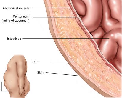 There are many different types of tissue that make up the abdominal wall and they each have their place in keeping the surgical abdomen closed. From deep to superficial, the anatomic layers that create the layers of abdominal wall are:
- Peritoneum
- Extraperitoneal fascia (deep fascia)
- Muscle
- Subcutaneous tissue (superficial fascia)
- Skin
It will depend on the location whether different layers are absent or present.
1. Peritoneum
Made up of two different layers, the peritoneum has one layer that positions itself in the cavity of the abdomen while the other one lines the area around the organs. Besides supporting the organs that are in the abdomen, the peritoneum lets the blood vessels, lymph vessels, and nerves pass over to the organs.
The visceral peritoneum actually covers the organs and the parietal peritoneum surrounds the wall of the abdomen and includes the organs. The peritoneal cavity can be found in between the two layers with a fine layer of fluid that keeps the peritoneal surfaces lubricated.
While the visceral peritoneum shares the blood that is used by the nerves and lymphatic vessels of the organs of the abdomen, the parietal peritoneum utilizes the nerve supply and circulation from the layers of abdomen wall.
2. Extraperitoneal Fascia (Deep Fascia)
Layers of abdominal wall include the extraperitoneal fascia whose amount and quality vary depending on where it is in the body. You will find that the paraneal fascia that is located around the kidneys can be fairly fatty and thick while the linea alba in the anterior can be fibrous and thin in the anterior of the wall of the abdomen.
3. Muscles
There are two groups of five muscles that are located in the wall of the abdomen. The groups consist of vertical muscles and flat muscles.
- The Flat Muscles
These muscles laterally flex and rotate the trunk. They can help to strengthen the abdominal wall and reduce the risk of herniation with their fibers running in different directions and crossing each other.
|
External Oblique |
This is the largest of the flat muscles that line the wall of the abdomen. The fibers meet in the middle and create a flat broad tendon. The aponeuroses of the flat muscles intertwine with each other and create the linea alba which runs from the sternum to the middle of the public bones. |
|
Internal Oblique |
While it is located right next to the external oblique, it is thinner and smaller in size. It has fibers that run perpendicular to the external oblique. At the midline point it creates fibers that contribute to the linea alba. |
|
Transversus Abdominis |
This one runs the deepest with its fibers running transversely. This muscle also contributes to the fibers of the linea alba and is close to the transversalis fascia. |
- The Vertical Muscles
Besides the flat muscles, there're also vertical muscles that run through the layers of abdominal wall. The two vertical muscles can be found along the middle of the body.
|
Rectus Abdominis |
This muscle is located on either side of the abdominal wall along the midline. It is divided in half by the linea alba. The strips of fibers that run through the muscle are what give people who work out a "six pack." The rectus abdominis keeps the pelvis stable while walking and keeps the ribs depressed. |
|
Pyramidalis |
This muscle is triangular in shape and found near the rectus abdominus. You can find the bottom of it on the pubis bone and the top is attached to the linea alba. |
4. Subcutaneous Tissue (Superficial Fascia)
This muscle is made up of connective tissue that is fatty. Its composition depends on where it is located.
If it is above the umbilicus, it is made up of a single sheet of tissue; if it is below the umbilicus, it has two layers – the superficial layer that is fatty and the deep layer which has a lot of membranes. There are superficial nerves and vessels that go between these two layers.
5. Skin
Skin is the outermost layer of the abdominal wall. It protects us from microbes and the elements, helps regulate body temperature, and permits the sensations of touch, heat, and cold.
Nerves That Run to the Muscles and Skin of Abdomen
In the layers of abdominal wall, there are a large number of nerves that go between the skin and muscles of the abdomen. They include:
- Thoracoabdominal nerves – There are five pair of these nerves and they run between the muscles of the abdominal wall to the muscles of the anterolateral wall of the abdomen. The cutaneous, lateral, and anterior branches bring a supply of nerves to the skin.
- Subcostal nerves – Located at the 12th thoracic spinal of the anterior rami, the nerves from this area run inferior from the 12th ribs down to the lower area of the navel. These nerves innervate the muscles of the abdominal wall and the skin that is located from the umbilicus to the iliac crests.
- Iliohypogastric nerves – These nerves run from spinal nerves of the first lumbar to create branches that go below the subcostals to the lower portion of the abdominal wall. They are responsible for innervating the skin that is over the iliac crests, inguinal areas, pubic areas and the regions that are underneath the navel.
- Ilioinguinal nerves - Located at the back of the first nerves of the lumbar spine, these nerves travel between the abdominal muscles to the inguinal canal. Their purpose is to innervate the skin on a man's scrotum and the area over the pubic bone in women, the labia majora and portions of the thigh. Other areas they oversee are the transversus abdominis and internal oblique muscles.
- Lateral cutaneous nerves on the thigh – Responsible for the skin on the anterolateral portions of the thighs, these nerves can be found running from the 2nd and 3rd nerves of the spinal lumbar to the thigh's iliacus muscles.
- Femoral nerves – Located between the 2nd and 4th lumbar in the spinal nerve area they run around the major muscles near the iliacus muscle as it goes towards the thigh so that the knee can extend properly.
- Obturator nerves – Located from the 2nd to the 4th spinal nerves of the lumbar, these nerves run through the major muscle of the psoas over to the pelvis area and into the thigh area where they are responsible for the abductor muscles.
- Lumbosacral trunk – Located around the 4th and 5th nerve roots of the lumbar, they cross over the sacrum and help to create the sacral plexus after descending on the pelvis.