
One of the complications of cancer is malignant pleural effusion, a condition where abnormal amounts of fluid collect between the two layers of tissue which lines between the outside of lungs (pleura) and the wall of chest cavity. This usually occurs in patients with advanced lung or breast cancer, accounting for up to 65% of its incidence. Other causes include lymphoma and pleural mesothelioma.
Symptoms of Malignant Pleural Effusion
Malignant pleural effusion causes uncomfortable symptoms like progressive shortness of breath and cough. The severity of these symptoms is often affected by your position, so that you may feel worse at certain positions. Leaning forward as well as lying on one side increases pressure on the lungs, which can cause pressure on the affected lung and intensify the symptoms.
Diagnosis of Malignant Pleural Effusion
The diagnosis consists of evaluation of clinical factors and laboratory tests. Symptoms that last for more than one month in the absence of fever may suggest the condition. Further assessment consists of tests such as:
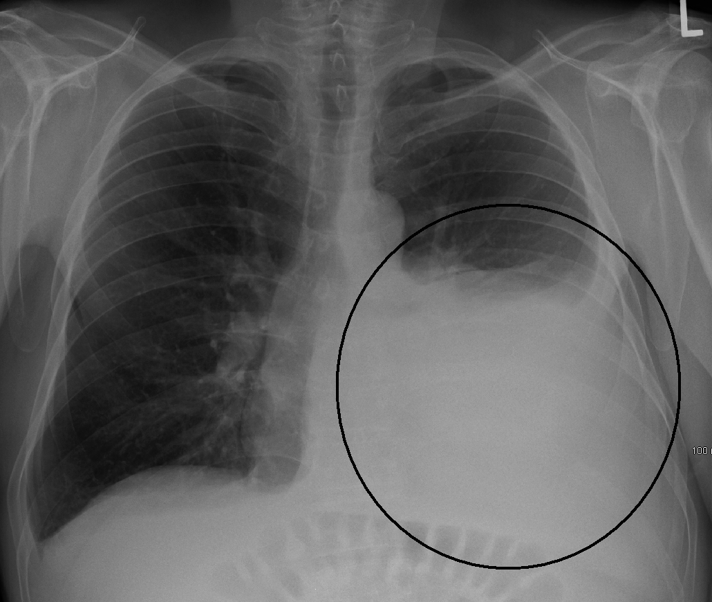
1. Chest Imaging
This helps to confirm the diagnosis of pleural effusion. The first test usually consists of a plain chest radiograph, which can also help demonstrate the underlying lung disease causing pleural effusion. Further evaluation using ultrasound can help distinguish malignant pleural effusion from other causes, since it is highly sensitive (73%) and specific (100%). This test can demonstrate the presence of pleural spread of the cancer (metastases) and thickening of the pleura that is greater than 1 cm. It can also reveal the presence of lymph nodes, thickening of the diaphragm to more than 7 mm, and a visible swirling pattern in the pleural fluid. These confirm the presence of pleural effusion that is probably caused by a malignancy.
2. Biochemical Analysis
Malignant pleural effusion is caused by fluids (exudates) produced by cancer cells. A low fluid pH is usually associated with poor response to treatment and survival.
3. Histopathology
Microscopic examination of the pleural fluid for cells (cytology) is positive in most cases. If doubtful, a pleural biopsy may be required. This may be done with the guidance of imaging and thoracoscopy for greater safety and sensitivity. Biopsy guided with a CT scan has 87% sensitive while blind needle biopsy only has less than 50% sensitivity.
4. Biomarkers
Malignant pleural effusion can be identified with the use of pleural fluid biomarkers and may be distinguished from other types of exudative effusion. These biomarkers that can exclude benign (non-cancerous) diseases like endostatin, vascular endothelial growth factor (VEGF), matrix metalloproteinases and other tumor markers like carcinoembryonic antigen. The biomarker called mesothelin has a greater sensitivity than cytology and a high specificity for the detection of malignant mesothelioma.
Treatments for Malignant Pleural Effusion
Since the presence of malignant pleural effusion indicates advanced cancer, the goal of treatment is palliative, which means reducing symptoms and improving the quality of life, but not to cure cancer. These treatments may include:
1. Thoracentesis
If the amount of fluid in the lungs is very small, doctors may leave it alone without treatment. However, if it causes severe symptoms, the fluid may be removed surgically by thoracentesis. However, the fluid frequently returns because cancer is still present.
2. Pleurodesis
Recurrent malignant pleural effusions often cause shortness of breath that can affect the patient's quality of life. Many patients improve after a procedure called pleurodesis. It involves inserting a tube into the space between the pleura and applying a substance like talcum powder between the membrane linings of the lungs. This results in inflammation which causes the two linings to fuse and prevents fluid accumulation in the pleural space.
3. Tunneled Pleural Catheter
Another common procedure is tunneling a catheter into the pleural space. It involves inserting a small tube, which is tunneled beneath the skin, into the pleural space. It leaves a small opening on your skin, which is covered with bandage. The catheter allows you to drain your own fluid (with a loved one's assistance) by using a vacuum container. This is usually indicated for recurrent malignant pleural effusion and is placed after thoracentesis. It helps relieve shortness of breath and makes you more comfortable.
4. Surgery
A persistent pleural effusion that does not respond to other techniques may be managed through surgery to drain fluids into the abdomen. This is called a pleurectomy. It involves removal of part of the pleura.
Another new medical treatment is called pleuroscopy. In some cases, chemotherapy may also help treat malignant pleural effusion that is due to some types of cancer like small cell lung cancer. However, it may not be effective in patients with non-small cell lung cancer.
Prognosis of Malignant Pleural Effusion
As previously mentioned, this condition often indicates the presence of advanced stage lung cancer or breast cancer. Patients in these stages often have a poor prognosis, with an average life expectancy of less than six months. Statistics show that the median survival time (50% have died and 50% still living) is four months.